 2017, Vol. 44
扩展功能

文章信息
- 瞿娇娇, 邹晓, 郁建平, 周家喜, 周叶鸣, 梁宗琦
- QU Jiao-Jiao, ZOU Xiao, YU Jian-Ping, ZHOU Jia-Xi, ZHOU Ye-Ming, LIANG Zong-Qi
- 被毛孢属两个昆虫病原真菌的重新描述
- Re-description of two entomopathogenic fungi in Hirsutella Pat.
- 微生物学通报, 2017, 44(11): 2615-2623
- Microbiology China, 2017, 44(11): 2615-2623
- DOI: 10.13344/j.microbiol.china.170020
-
文章历史
- 收稿日期: 2017-01-08
- 接受日期: 2017-03-03
- 优先数字出版日期(www.cnki.net): 2017-04-19


2. 贵州大学生命科学学院 真菌资源研究所 贵州 贵阳 550025;
3. 贵州大学 昆虫研究所 贵州 贵阳 550025
2. Institute of Fungal Resources, College of Life Sciences, Guizhou University, Guiyang, Guizhou 550025, China;
3. Institute of Entomology, Guizhou University, Guiyang, Guizhou 550025, China
被毛孢属(Hirsutella Pat.)是昆虫病原真菌中最丰富和最重要的类群之一,其无性型隶属于肉座菌目(Hypocreales)线形虫草科(Ophiocordycipitaceae)[1]。自1892年Patouillard[2]以嗜虫被毛孢Hirsutella entomophila Pat.作为模式种建立该属以来,该属真菌新成员不断被报道。被毛孢属真菌寄主广泛,在90个有效种内除H. uncinata Seifert & Boulay分离于哈克木属植物的蓇葖果[3]和H. piligena Bourdot & Galzin发现于毛发[4]外,寄生昆虫的有70余种,寄生线虫的有3种,寄生螨类的有12种,寄生蜘蛛的有3种[5]。该属能与其他无性型真菌区分的主要形态特征为:瓶梗细胞产孢,基部一般球形或柱状膨大,突然收缩成窄颈部,有再育现象,部分种类可呈扭曲状或螺旋状;分生孢子单孢、双孢或多孢,一般呈球形、椭圆形、梭形、橘瓣形或针形,孢子外常具有有色或无色黏液[6]。
被毛孢属真菌资源具有广阔的开发和利用价值。其中为人所熟知的是该类群真菌在螨类、茎线虫、根结线虫等植物病原线虫及一些鳞翅目昆虫的生物防治方面具有窄谱、专一性较好的防治效果[7-10]。另外,该属真菌中绝大多数类群都是线形虫草的无性阶段,而虫草是东南亚许多国家重要的传统药材,因此其生物活性物质和药用价值也被学术界和产业界所关注[11-12]。
被毛孢属的研究起源于20世纪20-50年代间,Petch等对被毛孢进行了大量研究,先后报道了25个新种,但当时发表的不少新种描述不够详细,缺乏形象的线条图及模式标本,而且原始文献多有损毁[13]。直到70–80年代,以Minter等为代表的菌物学者对被毛孢属真菌确立了现代科学定义[14-15]。21世纪以来,国内外对被毛孢属分类信息的报道相对较少,多为新种或亚种的报道。Simmons等对美国46种被毛孢进行了分子序列信息收集,并基于形态特征系统地对该属进行了系统发育研究,极大地丰富了该属分子遗传信息,但没有对分类信息缺失的种进行完善和补充[16]。
尽管当前真菌物种分类研究逐渐过渡到分子系统发育研究,但仅依靠DNA数据而不考虑其它因素(如生境信息、形态特征等)是不合理的,得到的将是一种单调乏味的序列相似性模式,而不是进化模式。形态学分类与分子学研究之间应该相互并存、相互补充,分子生物学技术将以全新的视角来弥补传统分类学的不足[17-19]。因此,基于传统形态分类特征对被毛孢的研究仍然具有重要的实际意义,利用分子系统学和形态鉴定相结合可以为Hirsutella属真菌综合分类及其系统发育提供更广阔的前景。目前,Hirsutella属仍有许多物种在公共数据库中的分类信息不完整或缺失,如主模式标本的遗失、原始描述中没有获得无性型菌株、没有提供用于现代分类标准的分子序列信息等,这都给后续研究带来极大困扰。因此,对Hirsutella属已知物种的重新描述和分子遗传学信息的增补是有必要的,这将为该类真菌的生物资源研究及后续开发利用奠定基础。
在对贵州省德江县及都匀市林区进行虫生真菌资源调查过程中,分别采集到两种寄生于沫蝉科(Cercopidae)一种成虫和松梢螟(Dioryctria splendidella Herrich-Schaeffer)成虫的真菌标本,对其进行了分离鉴定,分别为两个已知种:长白山被毛孢(H. changbeisanensis)和雷州被毛孢(H. leizhouensis)。由于这两个种的主模式标本及模式菌株均已遗失且原始分类信息都存在部分缺失,因此对这两种真菌进行了详细的形态描述、图解、培养性状和分子遗传学信息的补充。
1 材料与方法 1.1 材料 1.1.1 标本: 标本1采自贵州省德江县楠杆自然保护区(28°11′23″N,107°51′35″E),寄主为同翅目沫蝉科(Cercopidae,Homoptera)一种成虫。标本编号为GZUIFR-dj14,采集时间为2016年5月,菌株编号为GZUIFR-hir160527。标本与菌株保存于贵州大学真菌资源研究所(Institute of Fungal Resources Collection,Guizhou University,GZAC)。标本2采自贵州省都匀市马鞍山林场(26°15′46″N,107°30′52″E),寄主为鳞翅目螟蛾科松梢螟(Dioryctria splendidella,Pyralidae,Lepidoptera)。标本编号为GZUIFR-dy1,采集时间为2014年5月,菌株编号为GZUIFR-hir140506。标本与菌株保存于贵州大学真菌资源研究所(GZAC)。 1.1.2 培养基及主要试剂和仪器: PDA培养基参照文献[13]配制。葡萄糖、琼脂、苯胺蓝、乳酸、石炭酸、甘油及样本脱水试剂均为分析纯,国药集团化学试剂有限公司;真菌DNA提取试剂盒OMEGA®Fungal DNA kit,美国OMEGA Bio-Tek公司。光学显微镜BK5000,美国OPTEC公司;扫描电镜S-3400N,日本HITACHI公司。 1.1.3 染色液: 棉蓝染色液:苯胺蓝0.025 g,乳酸10.000 g,石炭酸10.000 g,甘油20.000 g,蒸馏水10 mL。配制完后用双层擦镜纸过滤后备用。 1.2 方法 1.2.1 无性阶段菌株分离与培养: 先将标本虫体用清水冲洗干净,用75%的乙醇消毒标本虫体表面3-5 s,掰断寄主露出内部组织,用接种针挑取组织块放在无菌、含有抗生素(链霉素40 U/mL,青霉素40 U/mL)的PDA平板上,挑取部位应靠近生前昆虫背部血体腔位置,在22下培养20 d后观察菌落特征并镜检微观形态。选取纯化的菌落保存编号,文中菌株编号分别为GZUIFR-hir160527和GZUIFR-hir140506。在PDA培养基上培养用于鉴定,培养条件为:光照12 h,黑暗12 h,22,20 d。 1.2.2 光学显微镜和扫描电镜观察: 用透明胶带黏取菌落边缘的菌丝,95%乙醇固定5 s后,用棉蓝染色3 min以上,光学显微镜下进行观察拍照。用于扫描电镜(Scanning electron microscope,SEM)观察的样品处理参照Cao等[20]描述的方法进行,略有改动,即从PDA培养基上挑取1 cm2左右的菌块,于4%戊二醛溶液中4浸泡过夜,用磷酸盐缓冲液(137 mmol/L NaCl,2.7 mmol/L KCl,8.1 mmol/L Na2HPO4,1.5 mmol/L KH2PO4,pH 7.4)冲洗3次,每次10 min;分别用30%、50%、70%、90%、95%、100%乙醇进行逐级脱水,每级10 min;脱水后使用超临界CO2干燥样品,最后喷金于扫描电镜下进行观察拍照。 1.2.3 DNA的提取、PCR扩增及测序: 从PDA平板上刮取菌丝0.05-0.10 g用于DNA的提取,采用真菌DNA提取试剂盒提取样本DNA。随后以真菌基因组DNA为模板,选用各引物对进行多基因序列的扩增,引物为:(1) 983F和2218R[21]用于tef1扩增;(2) LROR和LR5[22]用于扩增28S rRNA基因;(3) ITS1和ITS2[23]用于扩增ITS rDNA。PCR产物送上海Invitrogen公司纯化和测序。 1.2.4 基于多基因序列的系统发育分析: 测序结果经BioEdit软件分析和手工校正,经Sequin软件编辑后提交GenBank获取登录号;序列经Blast后下载相似性大于84%的相关近缘属菌株的tef1、ITS和28S rDNA 3个基因序列(表 1);序列进行多重比对并使用Sequence Matrix 1.7.8软件拼接,通过jMODELTEST 2.1.7运行合适的核苷酸置换模型[24];以Cordyceps gunnii Berk.为外群,用MrBayes 3.1.2重建系统发育树,运行50万代后舍弃25%的样本[25];最后使用FigTree 1.4.2编辑发育树图。| 种名 Species |
ITS rDNA | 28S rDNA | tef1 rDNA |
| H. leizhouensis | KY415573 | KY415580 | KY415587 |
| H. changbeisanensis | KY415578 | KY415586 | KY415592 |
| H. nodulosa | KM652165 | KM652117 | KM652000 |
| H. liboensis | KM652163 | KM652115 | KY415588 |
| H. leigongshanensis | KY415574 | KY415581 | KY415589 |
| H. satumaensis | KM652172 | KM652125 | KM652008 |
| H. tortricicola | KT368152 | KY415583 | KY415591 |
| H. lecaniicola | KM652162 | KM652114 | KM651998 |
| H. illustris | KM652160 | KM652112 | KM651996 |
| H. kirchneri | KM652161 | KM652113 | KM651997 |
| H. versicolor | KJ524678 | KM652150 | KM652029 |
| H. guyana | KM652158 | KM652111 | KM651994 |
| H. uncinata | KJ524691 | KJ524712 | - |
| H. subulata | KM652176 | KM652130 | KM652013 |
| H. necatrix | KM652164 | KM652116 | KM651999 |
| H. minnesotensis | DQ078757 | KJ524703 | - |
| H. gigantea | KJ524679 | KJ524700 | JX566980 |
| H. cf. haptospora | KM652166 | KM652118 | KM652001 |
| H. sinensis | KM652173 | KM652126 | KM652010 |
| H. strigosa | KM652175 | KM652129 | KM652011 |
| O. myrmicarum | JX566963 | JX566967 | JX566973 |
| H. thompsonii var. thompsonii | KM652177 | KM652131 | KM652014 |
| H. thompsonii var. vinacea | KM652194 | KM652149 | KM652028 |
| H. citriformis (Delphacidae) | KM652151 | KM652103 | KM651987 |
| H. citriformis (Cixiidae) | KM652153 | KM652105 | KM651989 |
| H. citriformis (Psyliidae) | KM652155 | KM652107 | KM651991 |
| H. thompsonii (Eriophyidae) | KM652179 | KM652133 | KM652016 |
| H. thompsonii (Tetranychidae) | KM652188 | KM652143 | KM652024 |
| H. thompsonii var. synnematosa | KM652193 | KM652148 | KM652027 |
| H. rhossiliensis (Heteroderide) | KM652168 | KM652121 | KM652004 |
| H. rhossiliensis (Criconematidae) | KM652170 | KM652123 | KM652006 |
| H. cryptosclerotium | KM652157 | KM652109 | KM651992 |
| Ophiocordyceps robertsii | KC561978 | EF468826 | EF468766 |
| O. stylophora | JN049828 | JN941449 | DQ522337 |
| O. acicularis | JN049820 | EF468805 | EF468744 |
| O. cf. acicularis | GU723765 | DQ518757 | DQ522326 |
| O. xuefengensis | KC631800 | KX090288 | KC631791 |
| O. cochlidiicola | AB027377 | KJ878884 | KJ878965 |
| O. sinensis | HM140631 | KU239985 | EF468767 |
| O. macroacicularis | AB968402 | AB968418 | AB968576 |
| Cordyceps gunnii | HM140630 | HM140633 | AY489616 |
| - | - | - | |
| 注:-:无序列. Note: -: No sequence. | |||

|
| 图 1 长白山被毛孢菌落特征和产孢细胞结构GZUIFR-hir160527 Figure 1 Colonies and conidiogenous cells structures of Hirsutella changbeisanensis GZUIFR-hir160527 注:A:表面被覆菌丝的沫蝉虫体;B、C:PDA培养基上培养20 d后形成的菌落,B为菌落正面,C为菌落背面;D、E、F:光学显微镜下的分生孢子和产孢细胞结构;G、H、I:扫描电镜下的产孢细胞和分生孢子.标尺:A为5 mm;B、C为10 mm;D-H为10 μm;I为5 μm. Note: A: The insect of Cercopidae coated with hyphae; B, C: Colonial morphology on PDA agar medium for 20 days culture, B for the positive of colony, C for the back of colony; D, E, F: Conidiogenous cells structure and conidia (LM); G, H, I: Conidiogenous cells and conidia (SEM). bar: A=5 mm; B, C=10 mm; D-H=10 μm; I=5 μm. |
|
|
虫体覆盖灰白色菌丝,无孢梗束产生。菌株在PDA培养基上生长缓慢,20-22培养20 d后菌落直径7.8 mm-13.0 mm;菌落表面隆起,呈不规则褶皱,边缘嵌入培养基,菌丝稀疏,呈绒毛状,正面灰白色,背面黑色,有褐色或黑色色素产生。菌丝透明、光滑,宽1.8 μm-3.5 μm;瓶梗直接着生于菌丝上,基部基本柱形,略膨大,(6.5-20.0) μm×(1.8-5.4)μm;向上变细长,(8.1-18.0)μm×1.0 μm,偶有再育产生2-3个小颈,光滑或具疣粒。分生孢子单孢,透明、光滑,拟卵形至橘瓣形,(4.0-7.0)μm×(2.5-3.5) μm;外被黏液,黏液层较薄,柠檬形至球形,(6.5-8.5)μm×(4.5-8.5) μm,一般含一个或两个孢子。
有性型:未知。
寄主:同翅目,叶蝉,沫蝉。
模式标本:GZUIFR-dj-14标本及其分离株GZUIFR-hir160527均保存于贵州大学真菌资源研究所保藏中心(GZAC)。
语义学:源于标本采集地。
MycoBank: MB 819473; GenBank: KY415578, KY415586, KY415592.
The insect covered with grey and white hyphae, without synnemata. The fungus spreading slowly on the PDA agar medium under 20-22 edded in the medium, much dark pigment secreting into the media make the back of colonies show black. Hyphae hyaline, smooth early, and wart found on the surface of old one, septate, ca. 1.8 μm-3.5 μm wide. Conidiogenous cell phialidic, mononematous, and borne perpendicular to the hyphae, basally inflated, cylindric, (6.5-20.0) μm×(1.8-5.4) μm long, tapering to a slender, verruculose, with narrow neck 8.1 μm-18.0 μm long, 1.0 μm wide; phialides formed directly from the mycelium end, single or occasional branched phialides with 2-3 in a whorl. Conidia ellipsoid or orange segment, (4.0-7.0)μm×(2.5-3.5) μm, usually embedded one or two in a conspicuous mucous sheath, (6.5-8.5) μm×(4.5-8.5) μm.
Teleomorph: Unknown.
Host: Homoptera, Cicadellidae; Cercopidae adult.
Holotypus: GZUIFR-dj14 and its asexual stage isolated strain GZUIFR-hir160527 were deposited in the GZAC.
Etymology: The species is named for the locality of the specimen.
2.2 雷州被毛孢[27]已知种 图 2 Hirsutella leizhouensis H.M.Fang & S.M.Tan Figure 2

|
| 图 2 雷州被毛孢菌落特征和产孢结构GZUIFR-hir140506 Figure 2 Colonies and conidiogenous cells structures of Hirsutella leizhouensis GZUIFR-hir140506 注:A:松枝上感染的松梢螟虫体;B、C:PDA培养基培养20 d后形成的菌落,B为菌落正面,C为菌落背面;D、E、F:光学显微镜下的分生孢子和产孢细胞结构.标尺:A为5 mm;B和C为10 mm;D、E、F为10 μm. Note: A: Infected Dioryctria splendidella specimens on a pine; B, C: Colonial morphology on PDA agar medium for 20 days culture, B for the positive of colony, C for the back of colony; D, E, F: Conidiogenous structure and conidia (LM). bar: A=5 mm; B, C=10 mm; D, E, F=10 μm. |
|
|
虫体表面被覆绒毛状白色菌丝,无孢梗束产生;该菌在PDA培养基上生长缓慢,20-22下20 d菌落直径为12 mm-16 mm;菌落正面奶白色,隆起,呈龟裂纹状纹沟,表面致密短绒毛状,背面为淡棕色或棕褐色,有色素渗透,扩散圈较大,1.0 cm-1.5 cm;菌落与培养基结合紧密,边缘培养基稍微下陷,背面无对应的培养基裂痕。菌丝无色,分枝,宽2.3 μm-3.0 μm,光滑或具疣。瓶梗直接着生于菌丝上,与菌丝呈直角或锐角,有的瓶梗为互生,有些菌丝顶端可直接形成瓶梗。瓶梗具有A、B两型:A型瓶梗呈瓶形,基部膨大,至中部突然变细成窄颈部,并呈螺旋状弯曲,长13.0μm-25.0 μm,基部宽1.8 μm-3.6 μm;B型瓶梗呈锥形,基部偶有横隔,上部逐渐尖削,呈螺旋状,长18.5 μm-30.0 μm,基部宽1.2μm-3.6 μm。瓶梗基部和颈部具细疣。瓶梗有再育现象,有1-3个分枝,最多不超过4个。分生孢子无色,呈椭球形或柠檬状,单生或双生,(2.0-3.6)μm×(3.6-5.4) μm;分生孢子具黏液。
有性阶段:未知。
寄主:鳞翅目,蠹蛾科,蔗褐蠹蛾;鳞翅目,螟蛾科,松梢螟。
模式标本:GZUIFR-dy-1标本及其分离株GZUIFR-hir140506均保存于贵州大学真菌资源研究所保藏中心(GZAC)。
语义学:源于标本采集地
MycoBank: MB355465; GenBank: KY415573, KY415580, KY415587.
The insect coated with villous and white hyphae, without synnemata. The fungus spreading slowly under 20-22 ℃ on the PDA agar medium and diameter to 12 mm-16 mm after 20 d; the colony is covered with irregular, dense bulges and creamy white flocculent aerial hyphae. The colonies margin is embedded in the medium, much light brown or brown pigment secreting into the media, the pigment diffusion circle larger, 1.0 cm-1.5 cm; the surface of the colony showed crack-like groove. Hyphae hyaline, smooth early and wart found on the surface of old one, septate, ca. 2.3 μm-3.0 μm wide. Phialides arise at direct or acute angle to the subtending hyphae and some of them opposite or directly formed at the top of hyphae. Two bottles of terrier type were formed, A-phialides, lageniform, middle part suddenly tapering to a twisted neck, width of base 1.8μm-3.6 μm, length 13.0 μm-25.0 μm; B-phialides, 18.5 μm-30.0 μm long, subulate with barely inflated base, 1.2 μm-3.6 μm broad, tapering to a slender, verruculose and twisted neck; Phialides single or occasional branched with 1-3 in a whorl. Conidia ellipsoid or lemon-like, (2.0-3.6) μm×(3.6-5.4) μm, usually embedded in mucous sheath.
Teleomorph: Unknown.
Host: Lepidoptera, Cossidae, Phragmatoecia castaneae Hübner; Pyraloidea, Dioryctria splendidella.
Typus: GZUIFR-dy1 and its asexual stage isolated strain GZUIFR-hir140506 were deposited in the GZAC.
Etymology: The species is named for the locality of the specimen.
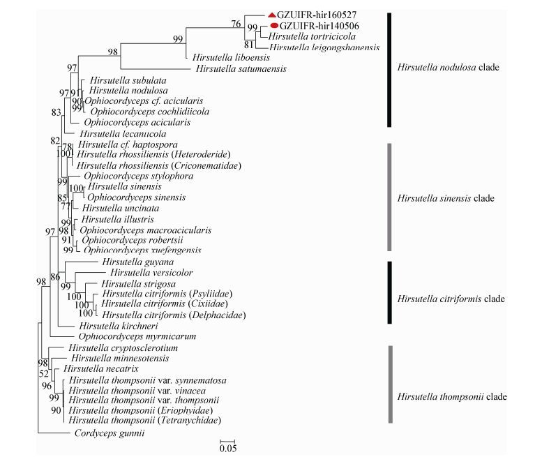
2.3 系统发育分析以古尼虫草Cordyceps gunnii Berk.作为外群构建的系统发育树(图 3)可大致分为4个类群:H. thompsonii、H. citriformis、H. sinensis和H. nodulosa,与Simmons等的研究结论基本一致[16]。其中H. leizhouensis与H. tortricicola关系密切并聚集在H. nodulosa分支中,这一类群的被毛孢形态学上的共同特征是瓶梗颈部具有螺旋或波浪结构,因此系统发育分析与形态学结构相吻合;H. leizhouensis与H. tortricicola之间的区别在于H. tortricicola只有一种产孢细胞,分生孢子橘瓣状,较细长,(2.7–3.6) μm×(1.4–1.8) μm;而H. leizhouensis产孢细胞双型,分生孢子柠檬状或卵圆形。H. changbeisanensis也聚到H. nodulosa分支中,但其bootstrap值只有76,并且该种具柱状或锥形瓶梗,颈部细长,分生孢子细长,与H. nodulosa分支具有明显差别。分子系统发育分析进一步印证了H. changbeisanensis和H. leizhouensis与其他物种的区别与联系。
|
| 图 3 菌株GZUIFR-hir160527和GZUIFR-hir140506及其相关种基于MrBayes分析tef1、ITS和28S rDNA 3个基因序列联合构建的系统发育树 Figure 3 Phylogenetic tree based on tef1, ITS, and 28S rDNA combined datasets of GZUIFR-hir160527 and GZUIFR-hir140506 related species 注:发育树节点的数值表示Bootstrap值;线段0.05表示序列差异的分支长度. Note: Number at notes present bootstrap percentages (based on 1 000 sampling); bar=5% represent sequences divergence. |
|
|
梁宗琦[26]首次描述报道了长白山被毛孢(H. changbeisanensis)新种,其寄主为同翅目的一种叶蝉,但仅简略描述了产孢结构形态,没有获得分离菌株及分子序列,并且主模式标本已经遗失。本文所描述的主模式标本寄主为同翅目沫蝉,显微观察确证了其产孢细胞基部基本柱形,略膨大,(6.5-20.0)μm×(1.8-5.4) μm,向上变细长,(8.1-18.0) μm×1.0 μm,与梁宗琦报道的形态基本一致,唯一的细微差别是本文研究菌株产孢细胞瓶颈具有明显的疣粒,可能是观察的时期不同所导致。被毛孢属中寄主为蝉科的种类较少,H. citriformis基部为球状膨大,颈部较长,超过20μm;H.longissima锥形瓶梗;H. atewensis柱状瓶梗,孢子梭形;H. heteropoda瓶梗极粗,颈部短小;H. floccosa瓶梗较长,超过20 μm;H. nivea孢子细长,(5.5-9.5) μm×0.5 μm;H. versicolor基部略膨大,颈部较宽,(8.0-12.0)μm×(2.5-3.0) μm,这些种均与标本菌株有明显差别。系统发育结果也表明该菌与其他被毛孢种能区分开来,与H. nodulosa分支有紧密联系,因此将该种定为H. changbeisanensis。
螺旋瓶颈是被毛孢属中一个特殊类群,目前在被毛孢属中共有10个种的产孢细胞颈部具螺旋或扭曲结构,这种结构的功能和进化意义还有待研究。本文描述标本寄主为松梢螟的被毛孢种也具有明显螺旋颈部,基于菌落形态、两型产孢细胞及螺旋瓶颈比对后其形态特征与雷州被毛孢(H. leizhouensis)基本一致。方焕谋等[27]首次报道了该种,寄主为蔗褐蠹蛾,并从标本中成功分离培养菌株,具两性瓶梗和螺旋瓶颈和详细的描述,但缺少分子遗传信息,模式标本已经遗失。文中补充了该被毛孢的新寄主、模式标本和无性型菌株及3个基因序列,系统发育分析显示该菌能很好地与H. nodulosa螺旋瓶颈分支聚在一起且有所区别。综合以上信息,将该种鉴定为H. leizhouensis。本文报道的新寄主为松梢螟,是松木的钻蛀性害虫,目前防治松梢螟的方法主要是抚育措施结合化学药剂防治,但防治效果不佳,生防制剂较少,H. leizhouensis的后续研究可用于马尾松等经济用林的生物防治。
| [1] |
Sung GH, Hywel-Jones NL, Sung JM, et al. Phylogenetic classification of Cordyceps and the clavicipitaceous fungi[J]. Studies in Mycology, 2007, 57: 5-59. DOI:10.3114/sim.2007.57.01 |
| [2] |
Patouillard N. Une Clavariée entomogène[J]. Revue Mycologique, 1892, 14: 67-70. |
| [3] |
Seifert KA, Boulay H. Hirsutella uncinata, a new hyphomycete from Australia[J]. Mycologia, 2004, 96(4): 929-934. DOI:10.1080/15572536.2005.11832940 |
| [4] |
Bourdot H, Galzin A. Hyménomycétes de France[M]. Sceaux, France: Marcel Bry, 1928, 142.
|
| [5] |
Ciancio A, Colagiero M, Rosso LC, et al. Phylogeny and morphology of Hirsutella tunicata sp. nov. (Ophiocordycipitaceae), a novel mite parasite from Peru[J]. Mycoscience, 2013, 54(5): 378-386. DOI:10.1016/j.myc.2013.01.002 |
| [6] |
Liang ZQ. Studies on classification of the genus Hirsutella Pat. Ⅰ. Advances and the characteristics of taxonomy[J]. Journal of Guizhou Agriculture College, 1990, 9(1): 58-68. 梁宗琦. 被毛孢属(Hirsutella Pat.进展及分类特征[J]. 贵州农院学报, 1990, 9(1): 58-68. |
| [7] |
Evans HC, Samson RA. Cordyceps species and their anamorphs pathogenic on ants (Formicidae) in tropical forest ecosystems I. The Cephalotes (Myrmicinae) complex[J]. Transactions of the British Mycological Society, 1982, 79(3): 431-453. DOI:10.1016/S0007-1536(82)80037-5 |
| [8] |
Cayrol JC, Castet R, Samson RA. Comparative activity of different Hirsutella species towards three plant parasitic nematodes[J]. Revue de Nématologie, 1986, 9(4): 412-414. |
| [9] |
Toledo AV, Simurro ME, Balatti PA. Morphological and molecular characterization of a fungus, Hirsutella sp., isolated from planthoppers and psocids in Argentina[J]. Journal of Insect Science, 2013, 13(1): 18. |
| [10] |
Hall DG, Hentz MG, Meyer JM, et al. Observations on the entomopathogenic fungus Hirsutella citriformis attacking adult Diaphorina citri (Hemiptera: Psyllidae) in a managed citrus grove[J]. BioControl, 2012, 57(5): 663-675. DOI:10.1007/s10526-012-9448-0 |
| [11] |
Thongtan J, Saenboonrueng J, Rachtawee P, et al. An antimalarial tetrapeptide from the entomopathogenic fungus Hirsutella sp. BCC 1528[J]. Journal of Natural Products, 2006, 69(4): 713-714. DOI:10.1021/np050549h |
| [12] |
Ge F, Gui L, Li WZ, et al. Study on effects of Hirsutella sinensis fermented mycelia on immunologic function in mice[J]. Chinese Journal of Clinical Pharmacology and Therapeutics, 2008, 13(8): 852-855. 葛飞, 桂琳, 李婉珍, 等. 中国被毛孢菌丝体对小鼠免疫功能的影响[J]. 中国临床药理学与治疗学, 2008, 13(8): 852-855. |
| [13] |
Zou X, Zhou JX, Liang ZQ, et al. Hirsutella shennongjiaensis, a new entomopathogenic species infecting Earwig (Dermaptera)[J]. Mycosystema, 2016, 35(9): 1070-1079. 邹晓, 周家喜, 梁宗琦, 等. 寄生革翅目蠼螋的被毛孢一新种[J]. 菌物学报, 2016, 35(9): 1070-1079. |
| [14] |
Minter DW, Brady BL. Mononematous species of Hirsutella[J]. Transactions of the British Mycological Society, 1980, 74(2): 271-282. DOI:10.1016/S0007-1536(80)80157-4 |
| [15] |
Evans HC, Samson RA. Cordyceps species and their anamorphs pathogenic on ants (Formicidae) in tropical forest ecosystems Ⅱ. The Camponotus (Formicinae) complex[J]. Transactions of the British Mycological Society, 1984, 82(1): 127-150. DOI:10.1016/S0007-1536(84)80219-3 |
| [16] |
Simmons DR, Kepler RM, Rehner SA, et al. Phylogeny of Hirsutella species (Ophiocordycipitaceae) from the USA: remedying the paucity of Hirsutella sequence data[J]. IMA Fungus, 2015, 6(2): 345-356. DOI:10.5598/imafungus.2015.06.02.06 |
| [17] |
Schindel DE, Miller SE. DNA barcoding a useful tool for taxonomists[J]. Nature, 2005, 435(7038): 17. |
| [18] |
Wang X, Huang B. Advancement of DNA barcoding in animal taxonomy[J]. Biotechnology Bulletin, 2006(4): 68-72. 王鑫, 黄兵. DNA条形编码技术在动物分类中的研究进展[J]. 生物技术通报, 2006(4): 68-72. |
| [19] |
Zhou JL, Zhao RL. Advances on DNA barcoding in fungi[J]. Microbiology China, 2013, 40(8): 1468-1477. 周均亮, 赵瑞琳. 真菌DNA条形码技术研究进展[J]. 微生物学通报, 2013, 40(8): 1468-1477. |
| [20] |
Cao YQ, Du MJ, Luo S, et al. Calcineurin modulates growth, stress tolerance, and virulence in Metarhizium acridum and its regulatory network[J]. Applied Microbiology and Biotechnology, 2014, 98(19): 8253-8265. DOI:10.1007/s00253-014-5876-3 |
| [21] |
Rehner SA, Buckley E. A Beauveria phylogeny inferred from nuclear ITS and EF1-α sequences: evidence for cryptic diversification and links to Cordyceps teleomorphs[J]. Mycologia, 2005, 97(1): 84-98. |
| [22] |
Rehner SA, Samuels GJ. Taxonomy and phylogeny of Gliocladium analysed from nuclear large subunit ribosomal DNA sequences[J]. Mycological Research, 1994, 98(6): 625-634. DOI:10.1016/S0953-7562(09)80409-7 |
| [23] |
White TJ, Bruns TD, Lee SB, et al. Amplification and direct sequencing of fungal ribosomal RNA genes for phylogenetics[M]//Innis MA, Gelfand DH, Sninsky JJ, et al. PCR Protocols: A Guide to Methods and Applications. San Diego: Academic Press, 1990: 315-322
|
| [24] |
Posada D. jModeltest: phylogenetic model averaging[J]. Molecular Biology and Evolution, 2008, 25(7): 1253-1256. DOI:10.1093/molbev/msn083 |
| [25] |
Ronquist F, Huelsenbeck JP. MrBayes 3: Bayesian phylogenetic inference under mixed models[J]. Bioinformatics, 2003, 19(12): 1572-1574. DOI:10.1093/bioinformatics/btg180 |
| [26] |
Liang ZQ. Two new entomopathogenic fungi of the genus HirsutellaPat[J]. Southwest China Journal of Agricultural Sciences, 1991, 4(1): 51-56. 梁宗琦. 被毛孢属的两种新昆虫病原真菌[J]. 西南农业学报, 1991, 4(1): 51-56. |
| [27] |
Fang HM, Tan SM. Hirsutella leizhouensis fang et tan[J]. Acta Mycologica Sinica, 1992, 11(1): 28-31. 方焕谋, 谭树明. 多毛孢属一个新种——雷州多毛孢[J]. 真菌学报, 1992, 11(1): 28-31. |