服务
文章信息
- 李晔, 陈振娅, 袁其朋
- Li Ye, Chen Zhenya, Yuan Qipeng
- 硫酸软骨素裂解酶ABC的研究进展
- Research progress in chondroitinase ABC
- 生物工程学报, 2015, 31(5): 621-633
- Chin J Biotech, 2015, 31(5): 621-633
- 10.13345/j.cjb.140459
-
文章历史
- Received: September 22, 2014
- Accepted: November 15, 2014
2. 北京化工大学生命科学与技术学院,北京 100029
2. College of Life Science and Technology, Beijing University of Chemical Technology, Beijing 100029, China
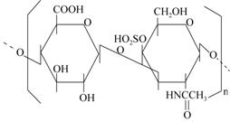
糖胺多糖作为蛋白聚糖的组成成分,主要分布在细胞外基质和细胞表面,用来指导很多生物过程,如细胞增殖、信号传输和炎症介导等[1, 2, 3]。硫酸软骨素 (Chondroitin sulfate,CS) 是糖胺多糖的一种,是以D-葡萄糖醛酸和2-乙酰氨基-2-脱氧硫酸-D-半乳糖通过β-1,3糖苷键相结合的双糖为基本单位,聚合而成的一类大分子多糖,双糖单位数目一般在50-70个,分子量约在5-50 kDa之间 (图1)。CS在软骨的细胞外间质中存在,并以O型糖肽键或N型糖肽键与核心蛋白的丝氨酸结合[4]。由于CS含有羧基和硫酸基,CS还是一种酸性多糖[5]。

|
| 图1 硫酸软骨素基本单元 Fig.1 The basic unit of CS. |
根据CS的结构及分子量的不同,将天然来源的CS分为A、B、C、D、E、F、H、K、L和M等[6]。常见的为CS A、B和C,CS A又称4-硫酸软骨素,硫酸基在氨基半乳糖的第4位C上 (图1)。CS B又称硫酸皮肤素,与CS A是异构体。CS C又称6-硫酸软骨素,硫酸基在氨基半乳糖的第6位C上[7, 8]。由于CS的分子量大,生物利用度低,在治疗脊柱损伤、轴突再生、抑制肿瘤发生等方面使得其很多功效不能得到有效的发挥,很多研究表明酶法制备低分子量的CS可以有效克服以上缺点,因此制备低分子量的CS具有重要意义[9, 10]。硫酸软骨素裂解酶 (Chondroitinase,ChSase) 是一类能将CS、软骨素、透明质酸等糖胺聚糖降解为不饱和二糖及寡糖的裂解酶[11]。目前,国内外有许多公司销售ChSase ABC,Sigma公司销售价格为414.18元/U,思清源公司销售价格为400元/U,主要用来检测CS含量。本文主要介绍了ChSase及ChSase ABC的分类和性质,ChSase ABC的晶体结构,ChSase ABC的分离纯化方法及重组菌的研究,分析了ChSase ABC作用、功效以及稳定性和固定化方面的研究进展,探讨并展望了ChSase ABC研究存在的问题及发展前景。
1 CHSase性质及分类ChSase是一类能将糖胺聚糖催化裂解为小分子多糖的酶[12],ChSase根据其作用底物的不同分为ChSase ABC、ChSase AC、ChSase B及ChSase C等类型,如表1所示。ChSase作为一种研究机体蛋白聚糖结构与功能的工具和一种新型药用酶日益受到人们的重视,2014年Makris等[13]将ChSase ABC、TGF-β1和赖氨酰氧化酶联合使用来治疗纤维软骨损坏后的功能修复,结果发现界面抗拉刚度和强度较未经ChSase ABC处理的软骨组织分别增加了730%和745%。Chen等[14]发现ChSase ABC可以使脑梗死后的神经再生,同时还可以抑制脑梗死的发生。Siebert等[15]发现ChSase ABC在治疗脊柱损伤过程中,可以显著提高轴突再生。
| Enzyme | Substrate | EC |
| ChSase AC | CS A, CS C, Hyaluronic acid (HA) | [EC 4.2.2.5] |
| ChSase B | CS B, HA | - |
| ChSase C | CS C, HA | - |
| ChSase ABC | CS A, CS B, CS C, HA | [EC 4.2.2.4] |
肝素黄杆菌Pedobacter heparinus中的ChSase AC和ChSase B酶活性和底物特异性已经被阐明[16, 17]。ChSase B的晶体结构和共晶结构表明了它与底物的结合位点,ChSase B也是钙依赖的裂解酶[16, 18, 19]。ChSase AC的晶体结构表明,其有与底物结合的特殊位点[17]。由于ChSase C的作用底物有限,目前有关ChSase C的报道还很少。
2 ChSase ABC简介硫酸软骨素裂解酶ABC (Chondrotinase ABC,ChSase ABC) 是研究糖胺多糖结构的工具酶[20]。普通变形杆菌Proteus vulgaris是ChSase ABC的主要来源,ChSase ABC分为ChSase ABCⅠ和ChSase ABCⅡ(EC 4.2.2.4)。两种酶对相同的底物有不同的作用位点,催化时具有不同的动力学参数[11, 21, 22, 23]。这两种酶已经在先前的报道中被纯化和研究。Prabhakar等[22, 23]克隆得到ChSase ABCⅠ和ChSase ABCⅡ的编码基因,并成功在大肠杆菌中表达,同时测定了该酶的活性和最佳反应条件。笔者所在的实验室从P. vulgaris (KCTC 2579) 中成功克隆到ChSase ABCⅠ的基因序列,在Escherichia coli中表达,获得可溶性的ChSase ABCⅠ,并对重组酶进行分离纯化,后对其酶活、比酶活及动力学常数等进行了测定[24]。
2.1 ChSase ABCⅠ和Ⅱ的基本性质ChSase ABCⅠ和ChSase ABCⅡ的分子量,分别为100 kDa和105 kDa[21],ChSase ABCⅠ和ChSase ABCⅡ都含有大量Asn、Gln和Leu,两个酶的N端氨基酸主要由Ala和Leu构成。DNA结构分析表明,ChSase ABCⅠ基因含有 3 063 bp的开放阅读框架,其中含有72 bp的前导肽序列,编码成熟后将切去24个氨基酸的前导肽序列[25],ChSase ABCⅡ基因含有2 973 bp的开放阅读框。
酶学基本性质显示,当以CS C为底物,ChSase ABCⅠ测定温度37 ℃,缓冲液为40 mmol/L Tris-HCl (pH 8.0),ChSase ABCⅡ测定温度40 ℃,缓冲液与ChSase ABCⅠ相同,ChSase ABCⅠ催化效率比ChSase ABCⅡ高,ChSase ABCⅠ的Km比ChSase ABCⅡ低,Vmax比ChSase ABCⅡ高[21],具体数据如表2所示。
| ChSase ABCⅠ | ChSase ABCⅡ | |
| Optimum temperature (℃) | 37 | 40 |
| Optimum pH | 8.0 | 8.0 |
| Km (μmol/L) | 66 | 80 |
| Vmax (mmol/(L·min·mg protein)) | 310 | 34 |
研究表明,对于不同的底物,测定温度为37 ℃,缓冲液为40 mmol/L Tris-HCl (pH 8.0),以CS A为底物时,ChSase ABCⅠ比酶活为400 U/mg,ChSase ABCⅡ比酶活为25 U/mg。以CS B为 底物时,ChSase ABCⅠ比酶活为180 U/mg,ChSase ABCⅡ比酶活为23 U/mg。以透明质酸为底物时,ChSase ABCⅠ比酶活为3 U/mg,ChSase ABCⅡ比酶活为5 U/mg。因此,对于不同的底物,ChSase ABCⅠ和ChSase ABCⅡ的催化活性有显著的差异。总体来说,ChSase ABCⅡ催化活性比ChSase ABCⅠ低[21]。笔者所在的实验室比较来源于P. vulgaris的ChSase ABCⅠ和ChSase ABCⅡ的酶活及比酶活,发现在一定实验条件下,ChSase ABCⅠ的总酶活达到3 180 U/L发酵液,与国内报道的最高水平2 000 U/L发酵液[26]相比高出1 180 U/L发酵液,发酵液经过分离纯化后比酶活为76 U/mg,纯度达到90%。而相同条件下检测不到ChSase ABCⅡ的酶活,该结果与ChSase ABCⅡ在P. vulgaris中的表达量及催化活性都比ChSase ABCⅠ低的结论相符[24]。
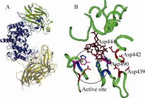
2.2 ChSase ABCⅠ和Ⅱ的晶体结构ChSase ABCⅠ由997个氨基酸残基构成,ChSase ABCⅡ由990个氨基酸残基构成[21]。通过重原子置换法 (Multiple isomorphous replacement,MIR) 和多波长反常散射法 (Multi-wavelengthanomalous diffration,MAD) 技术分析,ChSase ABCⅠ在1.9Å的分辨率下R因子和自由R因子分别为0.157和0.214,具有很好的立体化学性,晶体结构如图2A所示[27]。ChSase ABCⅠ的N端是由1-210位的氨基酸构成的双层β折叠和一个短的α螺旋,该区域的氨基酸序列与裂解碳水化合物酶相似,如木聚糖酶和一些凝集素。中间区域是由211-593位氨基酸构成的15个α螺旋,是主要的催化区域。C端由594-997位氨基酸构成的4个反向平行的β折叠,氨基酸序列与ChSase AC酶的C端有21%的相似性,与肺炎链球菌Streptococcus pneumoniae的透明质酸酶有 17 %-19 %的相似性,ChSase ABCⅠ的C端氨基酸的主要作用是裂解CS A、CS C和透明质酸[27, 28]。
电子密度图显示,ChSase ABCⅠ在中间的α螺旋区域495-530个氨基酸的部位有活性位点,与ChSase AC结构对比后,推测其活性位点为His501、Tyr508和Arg560,并通过定点突变的方法对其进行了验证[22, 29]。结果表明,His501、Tyr508和Arg560都是酶发生催化反应过程中的重要位点,其中Tyr508在裂解CS A、CS C和透明质酸过程中起着重要作用[27, 29, 30]。笔者所在的实验室已成功通过定点突变的方法验证了His501、Tyr508和Arg560为其催化反应的活性位点。2006年Prabhakar等[31]基于钙离子结合机制推测了ChSase ABCⅠ的裂解机理,通过定点突变和结构模拟分析,发现Asp442、Asp444和Tyr392是与钙离子距离最近的氨基酸,钙离子与上述位点结合,朱红色的四糖底物 (图2B) 与ChSase ABCⅠ以完全开放的方式结合后,His501、Tyr508和Arg560发挥裂解作用,通过β消除作用将二糖重复单元之间的β-1,3糖苷键切断,降解产物为寡糖,主要是不饱和二糖[32]。
ChSase ABCⅡ的晶体结构与ChSase ABCⅠ非常相似,主要包含3个主要的区域,N端是由14-170位的氨基酸构成的双层β折叠,且Ca2+结合在第2和第3个β链之间。中间区域是由171-593位氨基酸构成的双层的 (α/α)5 折叠,与ChSase AC,透明质酸裂解酶,黄原胶裂解酶和肝素酶Ⅱ相似,有一个与底物结合区域。C端由594-1014位氨基酸构成的4个反向平行的β折叠[27, 33]。与ChSase AC结构对比后,推测其活性位点为His454、Tyr461、Arg514和Glu628,同样通过定点突变实验验证了推测结果[33, 34]。
3 ChSase ABC的发酵生产ChSase ABC最早的生产方法是从原始菌中提取获得,国内最早分离得到的产ChSase ABC的菌株如表3所示,国外的研究重点在ChSase ABC的重组表达方面。
| Strains | MW (kDa) | Optimum temperature (℃) | Optimum pH | Results | References |
| P. vulgaris 6010 | - | - | - | The specific activity was improved by 91 times | [20] |
| Sphingomonas paucimobilis | 82.3 | 40 | 6.5 | The enzyme activity of exoenzyme was 2 U/mL | [26] |
| Proteus penneri | - | 30 | 7.5 | The enzyme activity was 0.3 U/mL | [12] |
| Aeromonas sobria | 80 | 37 | 7.0 | The enzyme activity was 0.9 U/mL | [35] |
国内对于ChSase ABC的发酵生产研究有很多,但是存在许多问题。一方面发酵生产ChSase ABC的菌株多数为胞内生产方式[22, 27, 29, 31],且得到的酶活偏低。胞内酶要经过细胞破碎才能得到ChSase ABC,破碎过程中存在细胞破碎不完全、酶活损失、消耗额外能源等弊端。因此发现和筛选出以分泌形式产ChSase ABC的菌株越来越重要,探索不同类型菌株,为后续ChSase ABC的重组表达研究提供重要信息。
另一方面,ChSase ABC发酵生产后的分离纯化也存在许多问题[20, 26]。分离纯化的步骤过于繁琐,能源消耗大,工业化成本太高,分离纯化后得到酶的比活力虽然有一定程度的提高,但总酶活在分离纯化的过程中损失了很多。因此,探索新型的分离纯化方法,对于ChSase ABC发酵生产显得尤为重要。另外,发酵条件的优化也是提高酶活的一个重要方面,研究发酵过程中,温度、pH、碳源、氮源、添加物等对发酵过程的影响,设计正交或响应面实验来优化发酵条件,从而提高产量和酶活。
4 重组ChSase ABC对于ChSase的重组表达研究主要集中在ChSase AC和ChSase B,吴敬君等[36]对Flavobacterium heparinum中的ChSase AC基因进行克隆,得到ChSase AC的基因cslA,通过与麦芽糖结合蛋白 (Maltose-binding protein,MBP) 构建融合表达载体,在大肠杆菌中表达,比酶活达到94.1 U/mg,且经过一步纯化后,ChSaseAC的纯度达到了95%。Pojasek[37, 38]对重组ChSase B进行了点突变,并对其应用进行了研究,同样在大肠杆菌中表达了肝素黄杆菌Flavobacterium heparinum的ChSase基因,并分离纯化得到了活性蛋白,当ChSase AC分别以CS A和CS B为底物检测时,Km分别为0.8 μmol/L和0.6 μmol/L,kcat分别为234 s-1和480 s-1,当ChSase B以CS B为底物检测时,Km和kcat分别为4.6 μmol/L和190 s-1。Tkalec等[39]在大肠杆菌中表达了ChSase AC和ChSase B,并在培养基中添加CS A作为碳源,且分离纯化得到活性蛋白,当ChSase AC以CS A为底物检测时,酶活为79.5 mU/(mL·A600unit),当ChSase B以CS B为底物检测时,酶活为29.4 mU/(mL·A600 unit)。
目前,对于ChSase ABC的重组表达研究很少,Prabhakar等[23, 29]将csl ABC重组到pET-28a载体中,表达后仅一部分蛋白以可溶形式存在,还有大部分以沉淀形式存在。高慧玲等[40]建立了ChSase ABCⅠ分泌型真核表达载体,并在胶质瘤细胞系中对其表达情况进行观察,结果ChSase ABCⅠ以分泌形式表达。1994年Nobuyuki等[25]对ChSase ABC进行克隆,并在大肠杆菌中表达,最后得到的重组酶与从P. vulgaris分离得到的一致。笔者所在的实验室对重组表达后的ChSase ABCⅠ,通过MBP柱纯化,并对其Km和kcat进行检测,分别为(73.1±4.1) μmol/L和(586.7±10.8) s-1,结果与Hamai等[21]直接从 P. vulgaris分离得到的ChSase ABCⅠ相近。
重组酶对采用亲和吸附法进行纯化 (如His-tag),存在许多弊端,成本高,吸附量有限,很难达到工业化生产的标准。
5 ChSase ABC的稳定性研究ChSase ABC应用于许多功能检测及功能验证实验,特别是在脊柱损伤治疗方面,ChSase ABC在进入大鼠体内后能否保持其原有的活性对于其应用前景尤为关键,因此ChSase ABC的热稳定性研究必不可少。2012年Mahdieh等[41]利用共溶剂的方法来提高ChSase ABCⅠ的热稳定性,选择了甘油、山梨醇和海藻糖3种化合物来影响ChSase ABCⅠ的热稳定性。结果表明,3种化合物添加后,ChSase ABCⅠ的催化活性和内荧光强度都有所提高,海藻糖的加入使得ChSase ABCⅠ的热稳定性得到了显著的提高,研究结果对临床用药方面的指导作用非常明显。2013年Mahdieh等[42]通过拉式图分析,Gln140的φ和ψ值均不是最佳,是造成热稳定性较差的主要因素,随后通过将Gln140突变成Ala、Gly和Asn来提高热稳定性。结果表明,分别在4 ℃、25 ℃和40 ℃,Gln140突变成Ala或Gly后,热稳定性和催化效率与野生型相比显著提高,而突变成Asn,热稳定性和催化效率明显下降。随着温度提高,酶的热稳定性逐渐降低,对于野生型,在4 ℃保存3周后酶活保持80%,在25 ℃保存2周后酶活完全丢失,在40 ℃保存25 min后酶活完全丢失。Gln140突变成Ala后,在4 ℃可以保存3周以上活性未改变,25 ℃时保存3周酶活降低到50 %左右,40 ℃时保存150 min后酶活完全丢失。张德盛等[43]为提高ChSase ABC的热稳定性,制备ChSase ABC-聚乳酸-聚乙醇酸共聚物缓释微球,并观察其体外释药特性,结果发现所制备的ChSase ABC微球形态均匀,粒径分布窄,再分散性好,释药平稳,3周内能维持有效的药物浓度。
ChSase ABC由于自身是蛋白质的原因,50 ℃时蛋白开始降解,是造成ChSase ABC不稳定的主要原因,限制了ChSase ABC在多方面的应用,尤其是在药用方面。目前ChSase ABC的稳定性研究还非常有限,还需要探索新型嗜热来源的酶及研究新型的稳定性制剂来保持酶的活性及稳定性。
6 ChSase ABC的固定化研究ChSase ABC在很多方面功效显著,因此对于ChSase ABC的需求量很大。大量生产的ChSase ABC如何重复利用及降低使用成本成了亟待解决的问题。酶的固定化技术是解决这一问题的重要方法,固定化酶是近十余年发展起来的酶应用技术,在工业生产、化学分析和医药等方面有诱人的应用前景。
酶的固定化方法有许多,常用的有吸附法、载体结合法、交联法、包埋法。ChSase ABC的固定化常用方法为吸附法和交联法。2004年蔡苏兰等[44]从温和气单孢菌YH311分离出ChSase ABC,然后利用海藻酸钠和纤维素固定化,固定化后ChSase ABC 的热稳定性及贮存稳定性得到大幅度的提高,固定化酶的收率为18.56%和18.86%。2010年牛海滨等[45]利用大孔树脂D380来固定化ChSase ABC,结果表明在最佳交联条件下,酶结合效率可达79.1%,固定化后酶Km达0.146 g/L,较游离酶高,具有较好的操作稳定性,适于工业化生产。2011年Huang等[46]将ChSase ABC固定在用壳聚糖和明胶做成的孔径为100-160 μm的神经导管上,且用聚乳酸做成直径为20-40 μm的微球,固定化之后酶活为0.07 U/mg,48 h后能保持48%的活性,聚乳酸微球在7 d后还能释放0.0162 U/mL的酶活。2013年Pakulska等[47]通过融合表达的方式,构建了C端有FLAG标签,N端有His和SH3标签的融合ChSase ABC (构建思路见图3),His用来纯化,同时和FLAG一起参与检测,SH3用来控制ChSase ABC释放,实验结果表明SH3已经成功固定化并控制ChSase ABC释放。
ChSase ABC的固定化技术还需进一步探索,寻找新型固定化酶的方法,既能保持酶的活性,也能达到重复利用的目的。
7 ChSase ABC 的应用ChSase ABC为特异性最为广泛的一类硫酸软骨素裂解酶,在基础研究中它用来构建二糖和寡糖的基因文库[48];研究糖胺多糖结构和性质[49];生产低分子量CS;作为CS的检测剂;在医药领域,用来治疗一些疾病,如脊柱损伤、轴突再生等[14, 15]。
7.1 降解CSCS作为药物有很多形式,硫酸软骨素片、硫酸软骨素注射液以及硫酸软骨滴眼液等,它有很多药效,如促进基质纤维的增长,改善血液循环,消除炎症,促进角膜愈合,改善眼部干燥等,它还具有抗凝血和防止血管硬化等活性,对冠心病、心肌梗塞等心血管病的防治有较好的疗效,对神经细胞、肾细胞等具有保护效应。尽管CS已有很多的药效,但小分子的CS药效更为显著。小分子的CS,以硫酸软骨素为主体构成的蛋白聚糖在软骨基质中起着一种“分子弹簧”的作用,具有止痛、促进软骨再生的功效,可从根本改善关节问题[32]。且低分子量的CS,无论口服或者注射用,吸收效果均优于高分子量CS[50]。低分子量的CS红外光谱性质、形貌特征等也都被探究,结果表明,低分子量的CS性能优于高分子量的CS[51, 52]。
7.2 检测CS目前药典[53]已明确表明,利用硫酸软骨素ChSase ABC来检测药物中CS的含量。鲍伦军等[54]用ChSase ABC酶解-高效液相色谱的方法检测鱼翅中的透明质酸,采用ZORBAX糖分析柱,紫外检测波长为226 nm,检测到鱼翅中透明质酸的质量分数为0.86%-1.96%。朱昱宁等[55]同样用酶解高效液相色谱 的方法检测CS的含量,采用Ultimate XB-NH2柱,紫外检测波长为232 nm,流动相为醋酸钠缓冲液 (pH 5.6)-乙腈 (950∶50),流速为1.0 mL/min,此色谱条件可将CS A、B和C分开。任丽萍等[56]用酶解液相色谱法建立了测定硫酸软骨素钠含量的方法,最低检测限为4.0 μg/mL,定量限为11.9 μg/mL.
7.3 医药领域的应用硫酸软骨素蛋白多糖 (CSPGs) 是中枢神经系统 (CNS) 损伤后胶质瘢痕的重要成分,具有抑制神经轴突再生的功效。CSPGs主要通过糖氨多糖链抑制神经再生,因此去除糖胺多糖或者干扰其合成均可使轴突再生,ChSase能够降解CSPGs,从而促进轴突再生[57]。除能促进轴突再生外,ChSase还具有缓解视网膜变形、提高后肢运动能力、降解囊性纤维变性部位的黏液物、抗肿瘤作用、增强软骨组织愈合等功效,具体如表4所示。
| Authors | Functions | References |
| Huang YM, et al | Degradation abnormal deposition CSPGs of rat’s retina, inhibition apoptosis of photoreceptor and promotion restoration of retina | [58] |
| Sun YX, et al | Improving movement ability of rat’s limb | [59] |
| Tester NJ, et al | Repairation spinal injury and improving movement ability of cat | [60] |
| Jiang FL, et al | Promotion growth of axons and shortening dominated time of target organs | [61] |
| Bhaskar KR, et al | Degradation mucus substance of diseased cells and improving function blocked organs | [62] |
| Brown MD, et al; Henderson N, et al; Kato F, et al | Degradation glycosaminoglycans, reduction pressure of spinal canal | [63−65] |
| Denholm EM, et al | Inhibition formation of melanoma and proliferation of tumor cells | [66] |
| Lee MC, et al | Promotion adhesion ability of chondrocytes and cartilage wound | [67] |
| Yao J | Preparation biodegradable scaffolds which can release ChSaseABC effectively and promotion repairation of rat’s spinal cord | [68] |
笔者所在实验室已成功克隆并在大肠杆菌中表达了来源于P. vulgaris (KCTC 2579) 的ChSase ABCⅠ,获得了目前最高的表达产量,并对重组蛋白进行了纯化,检测了纯化后酶的不同酶学性质及不同温度下的热稳定性[24]。目前重组菌构建越来越受到关注,ChSase ABC重组菌也越来越多,但是仍然面临着许多的困难:一方面通过菌体表达得到的重组酶,表达量不高,活性较低,大多数都以包涵体形式表达,再经过后续的包涵体分离纯化及复性,得到的酶量有限,活性也有损失。另一方面ChSase ABC的分离纯化,常用的是亲和吸附法,这种方法的成本很高,是制约酶工业化生产的主要瓶颈。另外,ChSase ABC酶的不稳定性也制约了该酶在诸如医药领域领域的应用。
ChSase ABCⅠ的发展受到上述诸多因素的限制,需从以下几方面入手来改变现状,首先,需要寻找新型菌株、表达策略及代谢途径来实现酶的高效分泌表达,避免后续的包涵体复性对酶活性的影响,实现其在工业化中的应用。其次,通过构建含有不同标签的融合表达载体来实现酶快速和高效分离纯化。最后,需研究新方法来增强酶的热稳定性,延长酶的保藏时间,如寻找耐热菌来源的ChSase ABCⅠ等。另外,ChSase ABCⅠ的固定化也是研究的一个重要方面,特别是在医药领域,如何找到能够承载ChSase ABCⅠ的合适的药物载体,来协助其完成特定的药效功能是亟待解决的问题。
| [1] | Bernfield M, Gotte M, Park PW, et al. Functions of cell surface heparan sulfate proteoglycans. Annu Rev Biochem, 1999, 68(1): 729-777. |
| [2] | Sugahara K, Mikami T, Uyama T, et al. Recent advances in the structural biology of chondroitin sulfate and dermatan sulfate. Curre Opin Struc Biol, 2003, 13(5): 612-620. |
| [3] | Bao X, Nishimura S, Mikami T, et al. Chondroitin sulfate/dermatan sulfate hybrid chains from embryonic pig brain, which contain a higher proportion of L-iduronic acid than those from adult pig brain, exhibit neuritogenic and growth factor binding activities. J Biol Chem, 2004, 279(11): 9765-9776. |
| [4] | Lamari FN, Karamanos NK. Structure of chondroitin sulfate. Adv Pharmacol, 2006, 53: 33-48. |
| [5] |
Ling PX, Chen L, Bian L. Progress on preparation technology of chondroitin sulfate. Food Drug, 2013, 15(1): 59-61 (in Chinese). 凌沛学, 陈磊, 边玲, 等. 硫酸软骨素制备工艺研究进展. 食品与药品, 2013, 15(1): 59-61. |
| [6] | Lauder RM. Chondroitin sulphate: a complex molecule with potential impacts on a wide range of biological systems. Compl Ther Med, 2009, 17(1): 56-62. |
| [7] |
Yu GL, Zhao X, Zhang TM. Structural character and quality control of chondroitin sulfate. Food Drug, 2010, 12(5): 153-157 (in Chinese). 于广利, 赵峡, 张天民. 硫酸软骨素的结构特点及其质量控制. 食品与药品, 2010, 12(5): 153-157. |
| [8] |
Xiong SL, Li AL, Wu ZM, et al. Extraction, separation and purification of chondroitin sulfate from chicken keel cartilage. Transa CSAE, 2009, 25(1): 271-275 (in Chinese). 熊双丽, 李安林, 吴照民, 等. 鸡胸软骨硫酸软骨素的提取及分离纯化. 农业工程学报, 2009, 25(1): 271-275. |
| [9] | Aich U, Shriver Z, Tharakaraman K. Competitive inhibition of heparinase by persulfonated glycosaminoglycans: a tool to detect heparin contamination. Anal Chem, 2011, 83(20): 7815-7822. |
| [10] | Garron ML, Cygler M. Structural and mechanistic classification of uronic acid-containing polysaccharide lyases. Glycobiology, 2010, 20(12): 1547-1573 |
| [11] | Yamagata T, Saito H, Habuchi O. Purification and properities of bacterial chondroitinase and chondrosulfatase. J Biol Chem, 1968, 243(7): 1523-1535. |
| [12] |
Tao K, Wang ZY, Guo JL, et al. The study of chondroitin sulfate lyase ABC bacteria screening and fermentation process. Chin J Antibiotics, 2004, 29(3): 138-141 (in Chinese). 陶科, 王忠彦, 国锦琳, 等. 硫酸软骨素裂解酶ABC产生菌的筛选及发酵工艺研究. 中国抗生素杂志, 2004, 29(3): 138-141. |
| [13] | Makris EA, MacBarb RF, Paschosa NK, et al. Combined use of chondroitinase-ABC, TGF-β1, and collagen crosslinking agent lysyl oxidase to engineer functional neotissues for fibrocartilage repair. Biomaterials, 2014, 35(25): 6787-6796. |
| [14] | Chen XR, Liao SJ, Ye LX, et al. Neuroprotective effect of chondroitinase ABC on primary and secondary brain injury after stroke in hypertensive rats. Brain Res, 2014, 1543: 324-333. |
| [15] | Siebert JR, Stelzner DJ, Osterhout DJ. Chondroitinase treatment following spinal contusion injury increases migration of oligodendrocyte progenitor cells. Exp Neurol, 2011, 231(1): 19-29. |
| [16] | Michel G, Pojasek K, Li Y, et al. The structure of chondroitin B lyase complexed with glycosaminoglycan oligosaccharides unravels a calcium-dependent catalytic machinery. J Biol Chem, 2004, 279(31): 32882-32896. |
| [17] | Huang W, Boju L, Tkalec L, et al. Active site of chondroitin AC lyase revealed by the structure of enzyme-oligosaccharide complexes and mutagenesis. Biochemistry, 2001, 40(8): 2359-2372. |
| [18] | Pojasek K, Raman R, Kiley P, et al. Biochemical characterization of the chondroitinase B active site. J Biol Chem, 2002, 277(34): 31179-31186. |
| [19] | Huang W, Matte A, Li Y, et al. Crystal structure of chondroitinase B from Flavobacterium heparinum and its complex with a disaccharide product at 1.7 Å resolution. J Mol Biol, 1999, 294(5): 1257-1269. |
| [20] |
Lin XS, Hou LZ, Yang TS. The preparation of chondroitin sulfate lyase ABC. J Norman Bethune Univ, 1997, 23(1): 104-105 (in Chinese). 林雪松, 侯立中, 杨同书. 硫酸软骨素裂解酶ABC的制备. 白求恩医科大学学报, 1997, 23(1): 104-105. |
| [21] | Hamai A, Hashimoto N, Mochizuki H, et al. Two distinct chondroitin sulfate ABC lyases: an endoeliminase yielding tetrasaccharides and an exoeliminase preferentially acting on oligosaccharides. J Biol Chem, 1997, 272(4): 9123-9130. |
| [22] | Prabhakar V, CapilaⅠ, Bosques CJ, et al. Biochemical characterization of the chondroitinase ABC I active site. Biochem J, 2005, 390(2): 395-405. |
| [23] | Prabhakar V, CapilaⅠ, Bosques CJ, et al. Recombinant expression, purification, and biochemical characterization of chondroitinase ABCⅡ from Proteus vulgaris. J Biol Chem, 2009, 284(2): 974-982. |
| [24] | Chen ZY, Li Y, Yuan QP. Expression, purification and thermostability of MBP-chondroitinase ABC I from Proteus vulgaris. Int J Biol Macromol, 2015, 72: 6-10. |
| [25] | Nobuyuki S, Masahiko S, Hiroshi N, et al. Cloning and expression in Escherichia coli of the gene encoding the Proteus vulgaris chondroitin ABC lyase. Appl Microbiol Biot, 1994, 41 (1): 39-46. |
| [26] |
Fu JY. Study of the screening, fermentation and purification of chondroitin sulfate enzyme[D]. Qingdao: Ocean University of China, 2012 (in Chinese). 付静芸. 产硫酸软骨素酶菌株的筛选、发酵、酶的分离纯化及性质的研究[D]. 青岛: 中国海洋大学, 2012. |
| [27] | Huang W, Lunin VV, Li Y, et al. Crystal structure of Proteus vulgaris chondroitin sulfate ABC lyase I at 1.9Å resolution. J Mol Biol, 2003, 328(3): 623-634. |
| [28] | Fethiere J, Eggimann B, Cygler M. Crystal structure of chondroitin AC lyase, a representative of a family of glycosaminoglycan degrading enzymes. J Mol Biol, 1999, 288(4): 635-647. |
| [29] | Prabhakar V, Capila I, Bosques CJ, et al. Chondroitinase ABC I from Proteus vulgaris: cloning, recombinant expression and active site identification. Biochem J, 2005, 386(1): 103-112. |
| [30] | Huang WJ, Matte A, Suzuki S, et al. Crystallization and preliminary X-ray analysis of chondroitin sulfate ABC lyasesⅠandⅡ from Proteus vulgaris. Acta Cryst, 2000, 56(7): 904-906. |
| [31] | Prabhakar V, Capila I, Raman R, et al. The catalytic machinery of chondroitinase ABC I utilizes a calcium coordination strategy to optimally process dermatan sulfate. Biochemistry, 2006, 45(37): 11130-11139. |
| [32] | Rye CS, Withers SG. Elucidation of the mechanism of polysaccharide by chondroitin AC lyase from Flavobacterium heparinum. J Am Chem Soc, 2002, 124(33): 9756-9767. |
| [33] | Shaya D, Hahn BS, Bjerkan TM, et al. Composite active site of chondroitin lyase ABC accepting both epimers of uronic acid. Glycobiology, 2008, 18(3): 270-277. |
| [34] | Shaya D, Hahn BS, Park NY, et al. Characterization of chondroitin sulfate lyase ABC from Bacteroides thetaiotaomicron WAL2926. Biochemistry, 2008, 47(25): 6650-6661. |
| [35] |
Yan HL, He HZ, Cai SL, et al. Screening and purification of chondroitinase from chondroitinase producing strains. Acta Microbiol Sin, 2004, 44(1): 79-82 (in Chinese). 阎浩林, 何汉洲, 蔡苏兰, 等. 硫酸软骨素酶产生菌的筛选及酶的分离纯化. 微生物学报, 2004, 44(1): 79-82. |
| [36] |
Wu JJ, Li Y, Zhang C, et al. Gene cloning and recombinant expression of chondroitinase AC from Pedobacter heparinus and characterization of recombinant fusion enzyme. Food Sci, 2013, 34(9): 127-134 (in Chinese). 吴敬君, 李晔, 张翀, 等. 肝素黄杆菌硫酸软骨素酶AC的高效重组表达体系构建及其酶学性质研究. 食品科学, 2013, 34(9): 127-134. |
| [37] | Pojasek K, Raman R, Sasisekharan R. Methods for purifying and isolation recombinant chondroitinases: US, 7129335. 2006-10-31. |
| [38] | Pojasek K, Shriver Z, Kiley P. Recombinant expression, purification, and kinetic characterization of chondroitinase AC and chondroitinase B from Flavobacterium heparinum. Biochem Bioph Res Co, 2001, 286(2): 343-351. |
| [39] | Tkalec LA, Fink D, Blain F. Isolation and expression in Escherichia coli of cslA and csl B, genes coding for the chondroitin sulfate-degrading enzymes chondroitinase AC and chondroitinase B, respectively, from Flavobacrerium heparium. Appl Environ Microb, 2000, 66(1): 29-35. |
| [40] |
Gao HL, Wang Q, Yu SZ, et al. The construction of a secretary eukaryotic expression plasmid of chondroitinase ABC I. Chin J Contemp Neurol Neurosurg, 2010, 10(4): 479-482 (in Chinese). 高慧玲, 王虔, 于士柱, 等. 硫酸软骨素酶ABC Ⅰ分泌型真核表达质粒的构建. 中国现代神经疾病杂志, 2010, 10(4): 479-482. |
| [41] | Mahdieh NR, Khosro K, Mahdi A, et al. Co-solvent mediated thermal stabilization of chondroitinase ABC I form Proteus vulgaris. Int J Biol Macromol, 2012, 50(3): 487-492. |
| [42] | Mahdieh NR, Khosro K, Mahdi A, et al. Enhancement of thermal stability of chondroitinase ABCⅠby site-directed mutagenesis: an insight from Ramachandran plot. Biochim Biophys Acta, 2013, 1834(2): 479-486. |
| [43] |
Zhang DS, Zhong DJ, Song YM, et al. Preparation and in vitro properties of controlled release poly (lactic-co-glycolic acid) microspheres incorporating chondroitinase ABC. J Clin Rehabil Tissue Eng Res, 2008, 12(32): 6247-6250 (in Chinese). 张德盛, 钟德君, 宋跃明, 等. 硫酸软骨素酶ABC-聚乳酸-聚乙醇酸共聚物缓释微球的制备及其体外性质. 中国组织工程研究与临床康复, 2008, 12(32): 6247-6250. |
| [44] |
Cai SL, Yan HL, He HZ. Purification and immobilization of chondroitinase from Aeromonas sobria YH 311. Chin J Biotech, 2004, 20(4): 584-588 (in Chinese). 蔡苏兰, 阎浩林, 何汉洲. 温和气单孢菌YH311硫酸软骨素裂解酶的分离纯化与固定化. 生物工程学报, 2004, 20(4): 584-588. |
| [45] |
Niu HB, Cai SL, Yan HL, et al. Immobilization of chondroitinase by Macroporous Resin D380. Chin J Biolog, 2010, 23(2): 203-207 (in Chinese). 牛海滨, 蔡苏兰, 阎浩林, 等. 大孔树脂D380固定化硫酸软骨素裂解酶. 中国生物制品学杂志, 2010, 23(2): 203-207. |
| [46] | Huang YC, Hsu SH, Chen MT, et al. Controlled release of chondroitinase ABC in chitosan-based scaffolds and PDLLA microspheres. Carbohyd Polym, 2011, 84(2): 788-793. |
| [47] | Pakulska MM, Vulic K, Shoichet MS. Affinity-based release of chondroitinase ABC from a modified methylcellulose hydrogel. J Control Release, 2013, 171(1): 11-16. |
| [48] | Michelacci YM, Horton DSPQ, Poblacion CA. Isolation and characterization of an induced chondroitinase ABC from Flavobacterium. Biochim Biophy Acta, 1987, 923(2): 291-301. |
| [49] |
Li YR. The Biochemistry and Research Methods of Extracellular Matrix. Beijing: People’s Medical Publishing House, 1988: 170-200 (in Chinese). 李玉瑞. 细胞外间质的生物化学及研究方法. 北京: 人民卫生出版社, 1988: 170-200. |
| [50] |
Xiong SL, Jin ZY. Preparation of chondroitin sulfate with different molecular weight and antioxitation capacity. Chin Tradit Patent Med, 2006, 28(9): 1343-1346 (in Chinese). 熊双丽, 金征宇. 不同分子量硫酸软骨素的制备和抗氧化活性探讨. 中成药, 2006, 28(9): 1343-1346. |
| [51] |
Shi MJ, Xiong SL, Wang YY, et al. Preparation and properties of low molecular weight chondroitin sulfate. Fine Chem, 2012, 29(11): 1088-1093 (in Chinese). 史敏娟, 熊双丽, 王莹莹, 等. 低相对分子质量硫酸软骨素的制备及其性质. 精细化工, 2012, 29(11): 1088-1093. |
| [52] | Xiao YL, Li PL, Cheng YN, et al. Enhancing the intestinal absorption of low molecular weight chondroitin sulfate by conjugation with α-linolenic acid and the transport mechanism of the conjugates. Int J Pharm, 2014, 465(1): 143-158. |
| [53] | National Pharmacopoeia Committee. Pharmacopoeia of People’s Republic of China. Part 2. Beijing: Chemical Industry Press, 2010: 984-986.国家药典委员会. 中华人民共和国药典. 2部. 北京: 化学工业出版社, 2010: 984-986. |
| [54] |
Bao LJ, Yang JC, He ZH, et al. Zymohydrolysis with chondroitinase ABC and high performance liquid chromatography used for the determination of hyaluronic acid in shark fin. Chin J Chromatogr, 2002, 20(6): 557-559 (in Chinese). 鲍伦军, 杨建成, 何振华, 等. 软骨素酶ABC酶解-高效液相色谱法测定鱼翅中的透明质酸. 色谱, 2002, 20(6): 557-559. |
| [55] |
Zhu YN, Ao L, Fu YH. Determination of chondroitin sulfate content by enzymatic HPLC. Chin J Biological Pharms, 2012, 33(6): 827-829 (in Chinese). 朱昱宁, 敖雷, 傅应华. 酶解高效液相色谱法测定硫酸软骨素的含量. 中国生化药物杂志, 2012, 33(6): 827-829. |
| [56] |
Ren LP, Yu HZ, Song YR, et al. Enzymolysis-HPLC determination of the content of chondroitin sulfate sodium. Chin J Pharm Analysis, 2012, 32(7): 1246-1248 (in Chinese). 任丽萍, 于海洲, 宋玉娟, 等. 酶解液相色谱法测定硫酸软骨素钠含量. 药物分析杂志, 2012, 32(7): 1246-1248. |
| [57] | Shields LB, Zhang YP, Burke DA, et al. Benefit of chondroitinase ABC on sensory axon regeneration in a laceration model of spinal cord injury in the rat. Surq Neurol, 2008, 69(6): 568-577. |
| [58] |
Huang YM, Gao PF, Xu YN, et al. Chondroitinase ABC alleviates photoreceptor apoptosis in the retinae . J Yangzhou Univ, 2012, 33(4): 19-23 (in Chinese). 黄玉苗, 高朋芬, 胥亚男, 等. 硫酸软骨素酶缓解视网膜变性大鼠光感受器细胞凋亡. 扬州大学学报, 2012, 33(4): 19-23. |
| [59] |
Sun YX, Liu N, Xu AH, et al. The effect of chondroitinase ABC combined with hyperbaric oxygen preconditioning treatment on the motor function of hindlimbs in spinal cord injury rats. Proc Anat Sci, 2012, 18(6): 526-529 (in Chinese). 孙永新, 刘宁, 徐爱华, 等. 硫酸软骨素酶ABC联合高压氧预处理对脊髓损伤大鼠后肢运动功能的影响. 解剖科学进展, 2012, 18(6): 526-529. |
| [60] | Tester NJ, Howland DR. Chondroitinase ABC improves basic and skilled locomotion in spinal cord injured cats. Exp Neurol, 2008, 209(2): 483-496. |
| [61] |
Jiang LF, Chen O, Chu TG. Decellularized nerve allograft treated with chondroitinase ABC for repair of peripheral nerve defects: an experimental study. Chin J Hand Surg, 2013, 29(4): 208-211 (in Chinese). 蒋良福, 陈鸥, 褚庭纲. 硫酸软骨素酶处理的异体去细胞神经修复周围神经缺损的实验研究. 中华手外科杂志, 2013, 29(4): 208-211. |
| [62] | Bhaskar KR, Lamont JT. Use of chondroitinase in the manufacture of a medicament in the treatment and prevention of muciod secretions: US, 1998046258. 1999-03-11. |
| [63] | Brown, MD. Method for treating intervertebral disc displacement with enzymes: US, 4696816. 1987-09-29. |
| [64] | Henderson N, Stanescu V, Cauchoix J. Nucleolysis of the rabbit intervertebral disc using chondroitinase ABC. Spine, 1991, 16(2): 203-208. |
| [65] | Kato F, Lwata H, Mimatsu K, et al. Experimental chemonucleolysis with chondroitinase ABC. Clin Orthop, 1990, 253(2): 301-308. |
| [66] | Denholm EM, Lin YQ, Silver PJ. Anti-tumor activities of chondroitinase AC and chondroitinase B: inhibition of angiogenesis, proliferation and invasion. Eur J Pharmacol, 2001, 416(3): 213-221. |
| [67] | Lee MC, Sung KL, Kurtis MS, et al. Adhesive force of chondrocytes to cartilage effects of chondroitinase ABC. Clin Orthop, 2000, 370(1): 286-294. |
| [68] |
Yao J. Controlled release tissue engineering scaffolds delivering chondroitinase ABC repair in the injury of spinal hemisection[D]. Ji’nan: Shandong University, 2013 (in Chinese). 姚军. 硫酸软骨素酶ABC缓释组织工程支架对大鼠脊髓半切损伤修复研究[D]. 济南: 山东大学, 2013. |
 2015, Vol. 31


